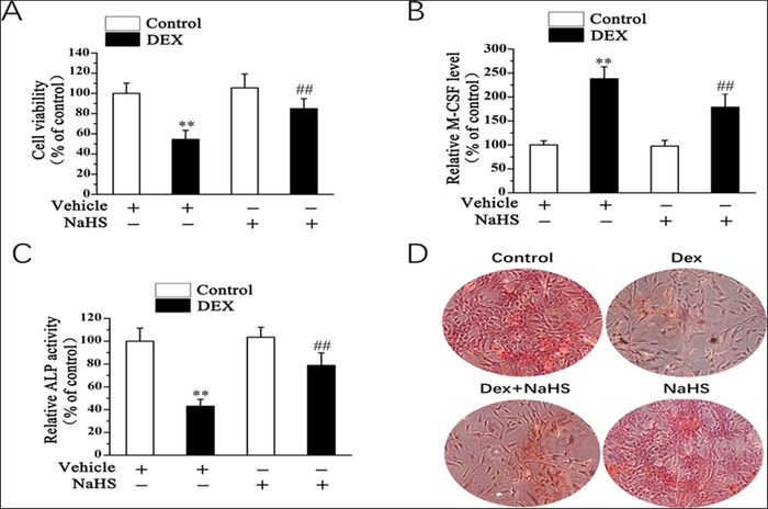
Figure 2.

NaHS mitigates DEX‐induced osteoblast dysfunction. 20 µM saline or NaHS was added to osteoblastic MC3T3‐E1 cells. Twenty‐four hours later, cells were subjected to the presence of 1 mM DEX for 48 H. Cell vitality (A), the level of M‐CSF (B), and ALP activity (C) were assessed. (D) As shown by ARS staining (day 14) (x40), NaHS attenuates DEX‐inhibited osteogenic differentiation in osteoblasts. All bar graphs represent means ± SEM (n = 4). **P < 0.01 versus control; ## P < 0.01 versus DEX.
