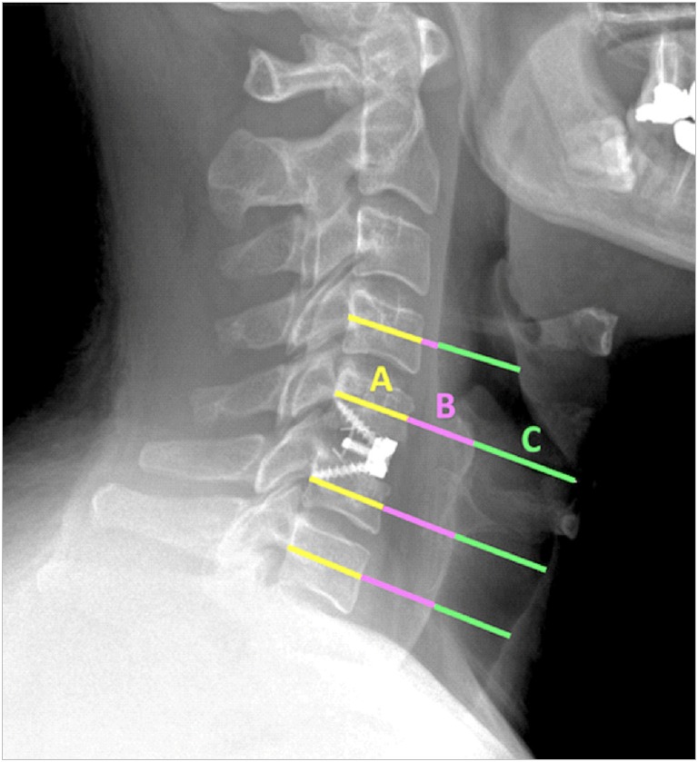
Fig. 1.

Postoperative lateral cervical radiograph of an anterior cervical discectomy and fusion patient who received a standalone cage demonstrates swelling and air index measurements at the operative level as well as one level above and below the fusion. All measurements are in the plane of the midvertebral body, parallel to the intervertebral discs. The measurements include anteriorposterior (AP) diameter of the vertebral body (A); prevertebral soft tissue swelling (B), from anterior cortex of the vertebral body to the posterior tracheal air window; and AP diameter of the tracheal air window (C).
