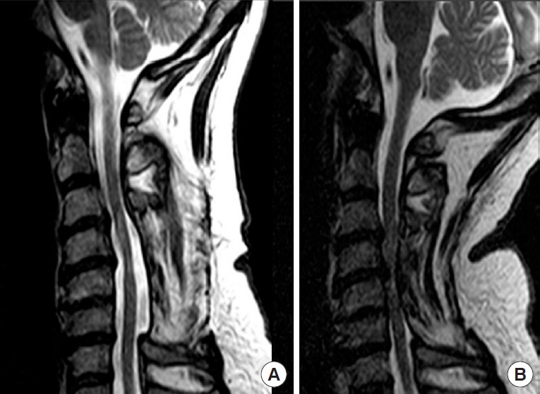
Fig. 1.

Sagittal T2 sequence cervical spine magnetic resonance imaging of an adult man who had a previous C3–6 laminectomy and late neurological deterioration after full recovery. In panel A, the neck is in neutral position and no evidence of spinal cord compression. In panel B, with the neck in extension, there is severe infolding of the posterior muscles and soft tissues into the spinal canal [11].
