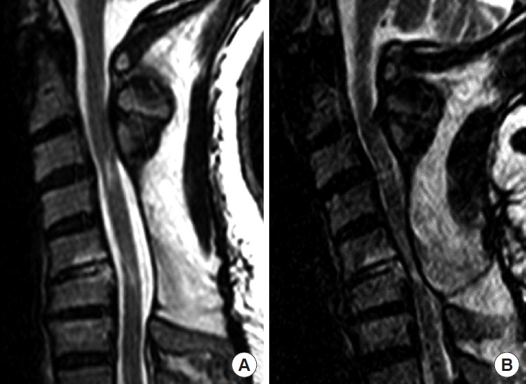
Fig. 2.

Sagittal T2 sequence cervical spine magnetic resonance imaging of a 56-year-old man who had a previous C4–6 laminectomy with fusion from C3–7 after rolling a vehicle. After surgery, he had no symptoms. Some months after the index surgery, he presented with increasing cervical pain and progression of cervical myelopathic symptoms when extending the neck. In panel A, the neck is neutral and no spinal cord is visualized. When the neck is extended, in panel B, there is severe spinal cord compression by the soft tissues in the back [11].
