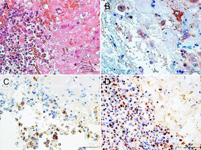
Figure 1.
Histopathological examination of the pituitary tumor. (A) Hematoxylin and eosin staining showing relatively large cells and pituitary adenoma cells adjacent and mixed within the boundary area. (B) Immunostaining for microtubule-associated protein 2 demonstrating positivity in large cells, indicating the presence of gangliocytoma. Immunostaining for growth hormone showing positivity but weak reactivity in pituitary adenoma cells (C) and CAM 5.2 immunostaining showing strong dot-like positive pattern (D), indicating the sparsely granulated subtype of pituitary somatotroph adenoma. Scale bar = 50 μm.

 This work is licensed under a