Figure 2.

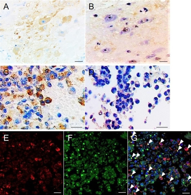
Immunohistochemical demonstration of growth hormone-releasing hormone (GHRH). Immunohistochemical staining using a pair of mirror section images showing no correspondence of microtubule-associated protein 2-positive (A) and GHRH-positive (B) cells, but demonstrating correspondence of growth hormone (GH)-positive (C) and GHRH-positive (D) cells. Double immunofluorescence staining for GH (E) and GHRH (F) revealing cells with colocalization (G). Arrowheads, colocalized cells. Scale bar = 50 μm in A–D; 20 μm in E–G.

 This work is licensed under a