Figure 1.

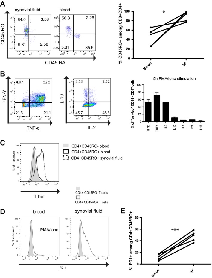
Synovial fluid (SF) T cells from patients with juvenile idiopathic arthritis (JIA) have a Th1 phenotype and express programmed death 1 (PD‐1). A, Left, Flow cytometric analysis indicating the frequencies of CD45RA+ and CD45RO+ cells among CD3+CD4+ T cells in synovial fluid and blood from patients with JIA. Results are representative of 5 experiments. Right, Percentage of CD3+CD4+ cells expressing CD45RO in blood and synovial fluid from patients with JIA (n = 5). * = P < 0.05 by 2‐tailed t‐test. B, Left, Expression of interferon‐γ (IFNγ), tumor necrosis factor (TNF), interleukin‐10 (IL‐10), and IL‐2 in ex vivo‐isolated synovial T cells after stimulation with phorbol 12‐myristate 13‐acetate (PMA) and ionomycin (iono), as analyzed by intracellular cytokine staining. Results are representative of 5 experiments. Right, Frequencies of synovial fluid CD4+ T cells expressing IFNγ, TNF, IL‐2, IL‐10, IL‐4, IL‐21, and IL‐17A after 5 hours of restimulation with PMA and ionomycin. Bars show the mean ± SEM (n = 5). C, T‐bet expression in CD4+CD45RO− and CD4+CD45RO+ T cells in blood and CD4+CD45RO+ T cells in synovial fluid from a patient with JIA. Results are representative of 3 experiments. D, PD‐1 expression in CD4+CD45RO− and CD4+CD45RO+ T cells isolated from blood stimulated with PMA and ionomycin and synovial fluid from a patient with JIA. Results are representative of 5 experiments. E, Percentage of CD4+CD45RO+ cells expressing PD‐1 in blood and synovial fluid from patients with JIA (n = 5). *** = P < 0.001 by 2‐tailed t‐test. Color figure can be viewed in the online issue, which is available at http://onlinelibrary.wiley.com/doi/10.1002/art.40939/abstract.
