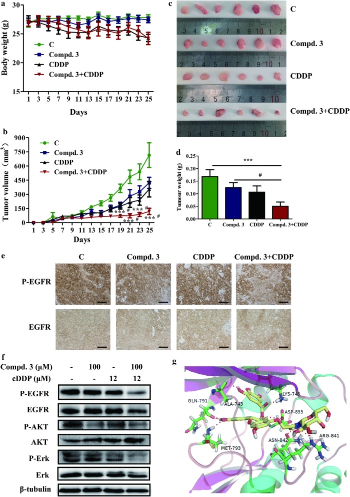
Fig. 7.
The antitumour effect of compound 3 and cDDP on NCI-H441 xenograft models. a The average body weight of each group. b The tumor volumes were measured every 2 days during the treatment. c, d The mice were sacrificed 25 days after drug treatment initiation, and the solid tumors were peeled from mouse subcutaneous tissue and tumor weights were measured. e Tumor tissues from NCI-H441 xenografts were immunostained with EGFR and P-EGFR antibodies. Magnification, ×20. Scale bar represents 100 μm. f Western blotting was used to assess EGFR, AKT, ERK, and their phosphorylated levels in tumor tissues. g Molecular docking model of compound 3 bound to EGFR (PDB code: 2ITY): ligand is colored by element type (C, yellow; O, red; N, blue; polar H, white), whereas key residues are shown as sticks (C, green; O, red; N, blue; polar H, white), and key interactions are denoted by a red-dotted line. Data represent the average of six independent mice per group (mean ± SD). ***p < 0.001 vs the control; #p < 0.05 cDDP + compound 3 vs compound 3

