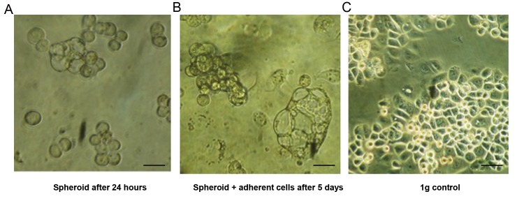
Fig.1.
Analysis of spheroid formation of breast cancer cells under simulated microgravity (0.003 g) after one and five days, under light microscopy. A. Formation of small spheroids can be observed after 24 hours, B. After 5 days, size of the cluster to tubular shaped spheroids increased, while part of the cells remained attached to the culture flask surface, and C. In the control group under 1 g conditions, the cells showed typical shape with flat morphology and rectangular to hexagonal borders (scale bar: 50 µm).

