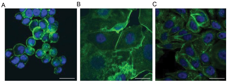
Fig.2.
Actin staining of breast cancer cells. A. In the spheroids under simulated microgravity, actin filaments arranged in a spherical shape with accentuation in the area of the cell membrane, B. In the adherent cells under simulated microgravity, spherical orientation beginning of the actin filaments was observed, and C. In the 1 g control group, actin filaments were arranged in a longitudinal manner with uniform distribution among the cytoplasm (scale bar: 25 µm).

