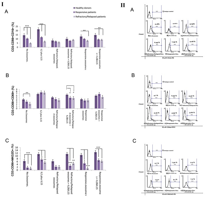
Fig.4.
Flow cytometer analysis of NK cell surface markers (CD16, NKG2D, and CD69) in the absence or presence of plasma-derived exosome of DLBCL patients. I. These surface markers were analyzed by gating on the live NK cells (CD56+CD3) of a representative DLBCL patient. A. NK cell labeled with PE-anti-human CD16 and PE Mouse IgG1, k Isotype control, B. NK cell labeled with FITC-anti-human CD69 and FITCI Mouse IgG1, k Isotype control, C. NK cell labeled with PE-anti-human CD314 (NKG2D) and PE Mouse IgG1, k Isotype control, i. Isotype control, ii. Unstimulated NK cell, iii. IL-2/ IL-15, iv. Plasma-derived exosomes of DLBCL refractory/ relapsed patients, v. Plasma-derived exosomes of DLBCL refractory/ relapsed patients plus IL-2/IL-15, vi. Plasma-derived exosome of responsive DLBCL patients and vii. Plasma-derived exosome of responsive DLBCL patients plus IL-2/IL-15. II. Average of the percentage of NK cells expressing A. CD16, B. CD69 and C. NKG2D was determined in each group (responsive DLBCL patients and refractory/relapsed DLBCL). Degree of significance was indicated by *P<0.05, **P<0.01, ***P<0.001. Each bar illustrates the mean ± SE. NK; Natural killer cells and DLBCL; Diffuse large B-cell lymphoma

