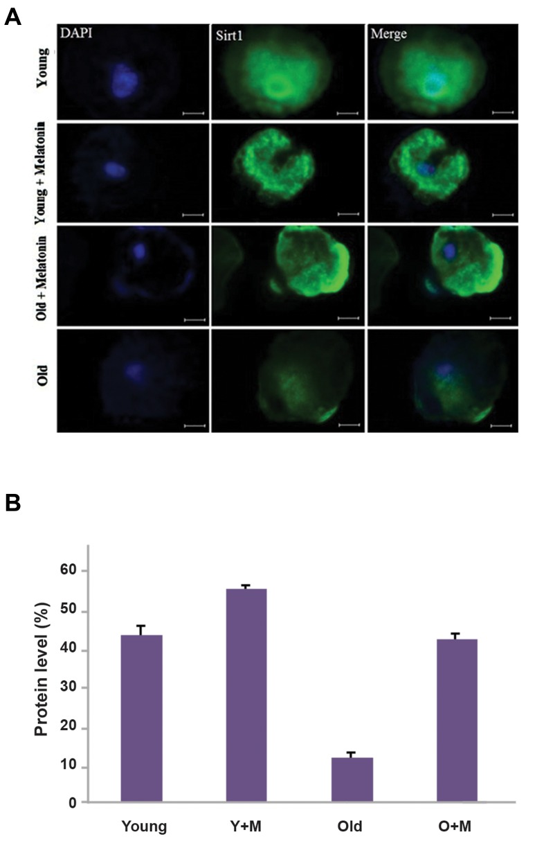
Fig.1.
The expression of SIRT-1 at the MII stage of in vitro matured oocytes, isolated from young and aged mice was evaluated usingimmunofluorescence staining. A. The micrograph represents the intensity of the SIRT-1 expression among the young MII oocyte, young MII oocyte+melatonin, aged MII oocyte+melatonin, and aged MII oocyte groups. The nuclei were stained by DAPI. The secondary antibody was conjugated with FITC and B. The expression of SIRT-1 in the aged MII oocyte+melatonin group was significantly higher than the aged MII oocyte (P<0.01). Accordingly, the SIRT-1 expression was elevated in the young MII oocyte+melatonin group compared with the young MII oocyte group (P<0.05) (magnification × 400, scale bars: 20 µm). Y+M; Young MII oocyte+melatonin and O+M; Aged MII oocyte+melatonin.

