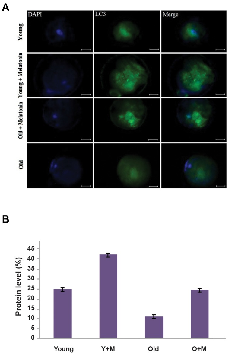
Fig.2.
The expression of the LC3 protein in in vitro matured MII oocytes, isolated from aged and young mice was determined by the Immunofluorescence staining. A. The micrograph represents a significant difference in intensity of the LC3 expression between the young MII oocyte, young MII oocyte+melatonin, aged MII oocyte+melatonin, and aged MII oocyte groups. The nuclei were stained by DAPI. The secondary antibody was conjugated with FITC (magnification ×400, scale bars: 20 µm) and B. Significantly higher levels of LC3 were found in the aged MII oocyte+melatonin compared with the aged MII oocyte groups (P<0.01). The expression of the LC3 was significantly higher in the young MII oocyte+melatonin than the young MII oocyte groups (P<0.01). Y+M; Young MII oocyte+melatonin and O+M; Aged MII oocyte+melatonin.

