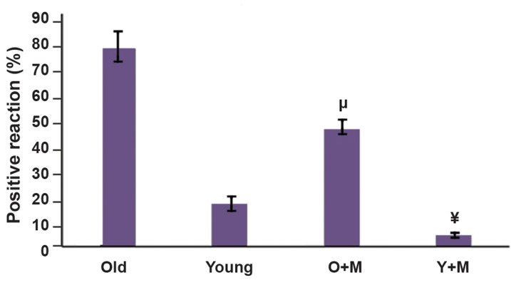
Fig.5.
Intracellular reactive oxygen specious (ROS) levels of MII in vitro matured oocytes were measured by immunofluorescence dye (DCFHDA) in all experimental groups, namely aged MII oocyte, young MIIoocyte, aged MII oocyte+10 µM melatonin, and young MII oocyte+10µM melatonin and they were quantified by the ImageJ software. Eachgroup consisted of 40-50 MII oocytes. The results were expressed asmean ± SD. The different symbols represent a significant differencebetween the two experimental groups. Although the ROS level wasdecreased in young MII oocyte+melatonin group compared with theyoung MII oocyte group, the difference was not statistically significant(4 ± 0.81 vs. 17 ± 3.09, P=0.71). The results also showed that there wasno significant difference between the aged MII oocytes+melatoninand young MII oocytes groups (P=0.10). ¥; P. 0.05 vs. aged group, µ; P<0.001 vs. young group, Y+M; Young+melatonin, and O+M; Aged+melatonin.

