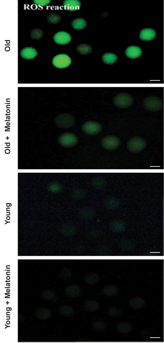
Fig.6.

The levels of DCFH-DA representing the reactive oxygen specious (ROS) production in MII in vitro matured oocytes, isolated from young and aged mice. The micrograph depicts the different intensity of ROS among the young MII oocytes, young MII oocytes+10 µM melatonin, aged MII oocytes, and aged MII oocytes+10 µM melatonin groups. The phase contrast of each group shows the morphology of oocytes. The fluorescence intensity of DCFH-DA was applied to probe ROS within the cytoplasm of oocytes (magnification: ×200, scale bars: 100 µm).
