Figure 4.

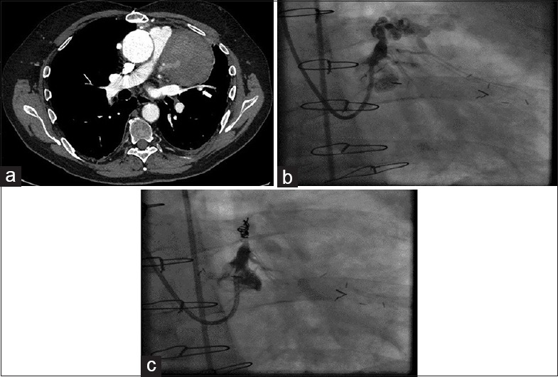
Computed tomography: (a) Computed tomogram showing a mediastinal mass with contrast extravasation (arrow) from the proximal left anterior descending aneurysm. (b) Right anterior oblique caudal view showing aneurysm in the proximal left anterior descending artery with extravasation of the contrast. (c) Right anterior oblique view showing closure of the aneurysm following coils insertion
