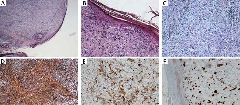
Figure 2.
A – Dome-shaped skin nodule composed of spindle cells arranged in a fascicular pattern. B – Atypical spindle cells arranged in a haphazard pattern, some with mitotic figures (arrows). No grenz zone of uninvolved dermis. C – Large, atypical, spindled, polymorphic cells with prominent nucleoli arranged in a haphazard pattern. D – Tumour cells were vimentin (mesenchymal marker) positive in immunohistochemical staining. E – Tumour cells were CD68 (histiocyte marker) positive in immunohistochemical staining. F – Ki67 index (proliferation index) was about 20% in tumour cells

