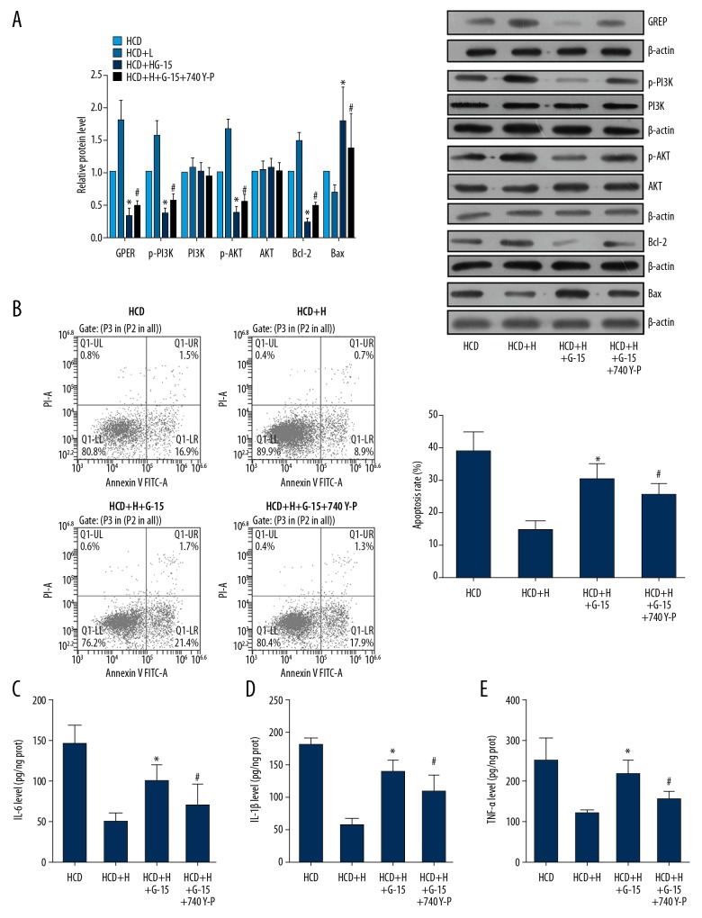
Figure 4.
Anti-AS function of Rb1 depended on the activation GPER- mediated PI3K/Akt pathway. ECs were isolated from AS and health rabbits and administrated with 80 μM Rb1, 100 nM G-15, and 25 mg/mL 740 Y-P for 24 h. (A) Representative images and quantitative analysis of western blotting detection of molecule expressions in GPER/PI3K/Akt axis. (B) Representative images and quantitative analysis of flow cytometry detection of apoptosis. (C) Quantitative analysis results of ELISA detection of blood IL-6. (D) Quantitative analysis results of ELISA detection of blood IL-1β. (E) Quantitative analysis results of ELISA detection of blood TNF-α. * P<0.05 vs. HCD+H group. # P<0.05 vs. HCD+H+G-15 group. Each assay was performed 5 times.

