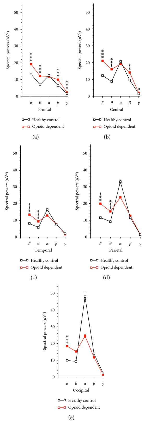
Figure 6.

Comparative analysis of the 5 areas. (a) Compared to the respective frequency bands in health controls, there were increases in powers of δ, θ, β, and γ oscillations, but not α oscillations of patients with opioid use disorder. ∗∗∗P < 0.001 vs. health controls determined by unpaired Student's t-test. (b) Compared to the respective frequency bands in health controls, there were increases in powers of δ and θ oscillations, but not α, β, or γ oscillations of patients with opioid use disorder. ∗∗∗P < 0.001 vs. health controls determined by unpaired Student's t-test. (c) Compared to the respective frequency bands in health controls, there were increases in powers of δ, θ, β, and γ oscillations, but not α oscillations of patients with opioid use disorder. ∗∗∗P < 0.001 vs. health controls determined by unpaired Student's t-test. (d) Compared to the respective frequency bands in health controls, there were increases in powers of δ and θ oscillations, but not α, β, or γ oscillations of patients with opioid use disorder. ∗∗∗P < 0.001 vs. health controls determined by unpaired Student's t-test. (e) Compared to the respective frequency bands in health controls, there were increases in powers of δ oscillations, but not θ, α, β, or γ oscillations of patients with opioid use disorder. ∗∗∗P < 0.001 vs. health controls determined by unpaired Student's t-test.
