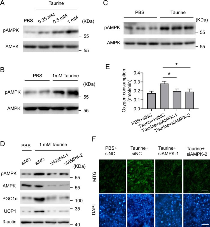
Figure 6.
Taurine-regulated PGC1α expression is AMPK signaling-dependent. A, C3H10T1/2 white adipocytes were treated with taurine at the indicated dose or with PBS as a control. After 24 h of treatment, cells were harvested for analyses. Cell lysates were subjected to Western blotting by using the indicated antibodies. B, fractionated and differentiated primary iWAT adipocytes were treated with taurine at a final concentration of 1 mm or with PBS as a control. After 24 h of treatment, cells were harvested for analyses. Cell lysates were subjected to Western blotting by using the indicated antibodies. C, mice were treated as indicated in Fig. 1. The tissue lysates of iWAT were subjected to Western blotting by using the indicated antibodies. D, C3H10T1/2 white adipocytes were transfected with the control siRNA (siNC) or two separate siRNAs (siAMPK1/2) against AMPKα1. 48 h post-transfection, cells were treated with taurine at a final concentration of 1 mm or with PBS as a control. After 24 h of treatment, cells were harvested for analyses. Cell lysates were subjected to Western blotting by using the indicated antibodies. E, cells were treated as in D, and OCR was measured. F, cells were treated as in D and then stained with MTG and DAPI. Representative images are shown. Scale bar, 100 μm. For statistical analysis, one-way analysis of variance plus Bonferroni's post hoc tests were carried out in E. All values are represented as means with error bars representing S.D. *, p < 0.05. n = 5 for each group.

