
Figure 2.
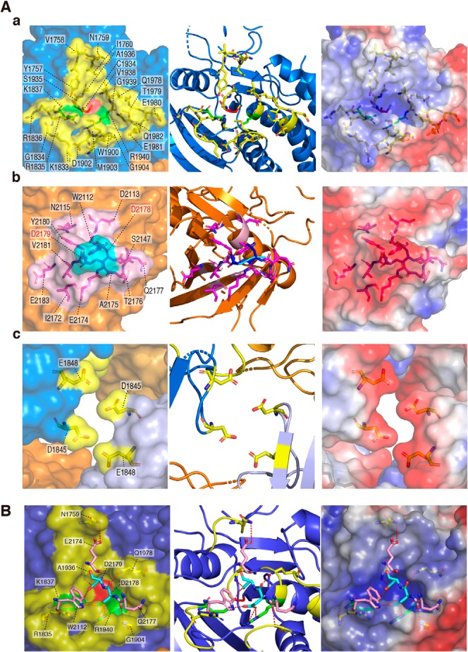
Dimer interface. A, a-c, amino acid residues forming the dimer interface are shown by a stick model inside the transparent surface (left), cartoon diagram (middle), and electrostatic potentials (right). Interface regions are colored as described in the legend to Fig. 1. Asp-2178 and Asp-2179 residues (shown in “red”) were mutated to Lys residues in DDKK mutants. B, interactions between the D1 and D2 dimer interfaces are shown by red dashed lines (details are given in Table S1).