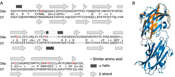
Figure 6.
Alignment of WztO9a-C (residues 270–423) to WztO7-C (residues 303–455). A, pairwise sequence alignment was performed by Blast (A). The indicated secondary structure elements are from the WztO9a-C crystal structure (Protein Data Bank entry 2R5O) and a Phyre2 model of WztO7-C. The residues coloured red are important for O-PS binding by WztO9a-C (23). B, cartoon representation overlay of WztO7-C model (orange) with WztO9a-C crystal structure (blue). The structural overlay was generated using PyMOL (Schrödinger, LLC).

