Figure 1.
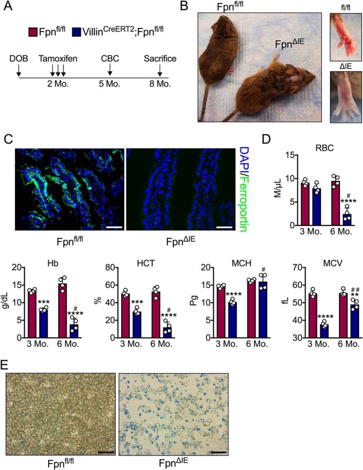
Intestinal epithelial ferroportin deletion in adult mice gives rise to progressive and end-stage iron deficiency anemia. A, schematic of the experimental design. Mo, month. DOB, date of birth; CBC, complete blood count. B, gross images of Fpnfl/fl and FpnΔIE mice 6 months after tamoxifen administration. C, representative ferroportin staining in duodenal sections of Fpnfl/fl and FpnΔIE mice; images at ×40, scale bars = 100 μm. DAPI, 4′,6-diamidino-2-phenylindole. D, analysis of RBCs, Hb, HCT, MCH, and MCV 3 and 6 months following tamoxifen injection. E, representative methylene blue staining for reticulocytes; images at ×60, scale bars = 150 μm. Mean ± S.E. are plotted. **, p < 0.01; ***, p < 0.001; ****, p < 0.0001 compared between Fpnfl/fl and FpnΔIE cohorts within each time point using two-tailed unpaired t test. #, p < 0.05; ##, p < 0.01 compared between individual FpnΔIE mice at the 3- and 6-month time points using two-tailed paired t test.

