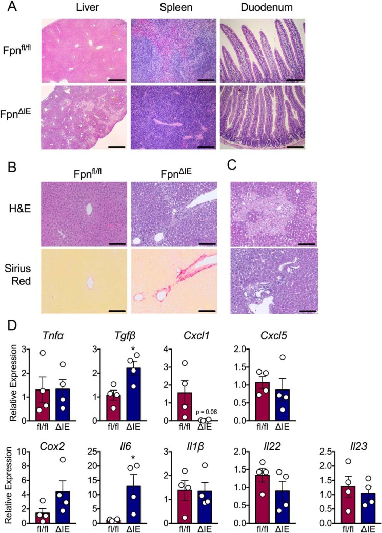
Figure 2.
Histological analysis of peripheral organs involved in iron homeostasis reveals inflammation and necrosis in the liver. A, representative H&E analysis of liver at ×5 (scale bars = 50 μm) and spleen and duodenum at ×20 (scale bars = 200 μm) from Fpnfl/fl and FpnΔIE cohorts. B, representative H&E and picrosirius red analysis of liver from Fpnfl/fl and FpnΔIE cohorts; images ×20, scale bars = 200 μm. C, additional representative H&E images of liver from FpnΔIE cohorts; images at ×20, scale bars = 200 μm. D, qPCR analysis of inflammatory genes in livers of Fpnfl/fl and FpnΔIE cohorts. All data are from mice 6 months post-tamoxifen treatment. Mean ± S.E. are plotted. Significance was determined using two-tailed unpaired t test. *, p < 0.5 compared between Fpnfl/fl and FpnΔIE cohorts.

