Figure 1.
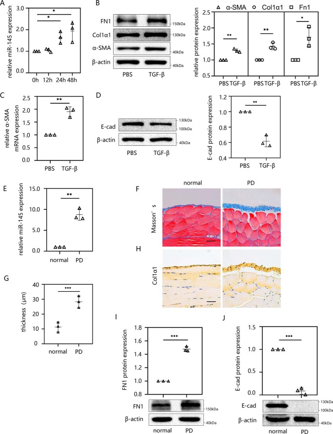
miR-145 is elevated in TGF-β1–induced EMT in vitro and PD-induced peritoneal fibrosis in vivo. The miR-145 expression level was determined by RT-qPCR. EMT and peritoneal fibrosis were detected by Masson's trichrome staining, immunohistochemistry, Western blotting, and RT-qPCR. A, miR-145 expression was examined in HMrSV5 cells treated with 10 ng/ml TGF-β1 for different times (0, 12, 24, and 48 h). B–D, HMrSV5 cells were starved for 12 h and then treated with 10 ng/ml TGF-β1. After 24 h, cells were harvested, and the protein levels of FN1, Col1α1, α-SMA (B), and E-cad (D) were evaluated by Western blotting. The mRNA level of α-SMA was evaluated by RT-qPCR (C). Male C57BL/6J mice were intraperitoneally injected daily with 3 ml of 4.25% dextrose PD solution for 4 weeks before the parietal and visceral peritoneum tissues were collected. E, the miR-145 expression level was analyzed by RT-qPCR. F and G, Masson's trichrome staining of anterior abdominal walls (F). G, thickness of the peritoneal membrane. H, immunohistochemistry was performed to analyze the expression of Col1α1 protein. I and J, Western blot analysis was used to detect the protein expression levels of FN1 (I) and E-cad (J). Each bar shows the mean ± S.D. (error bars) from three independent experiments. **, p < 0.01; ***, p < 0.001 versus the control group. Scale bar, 50 μm.

