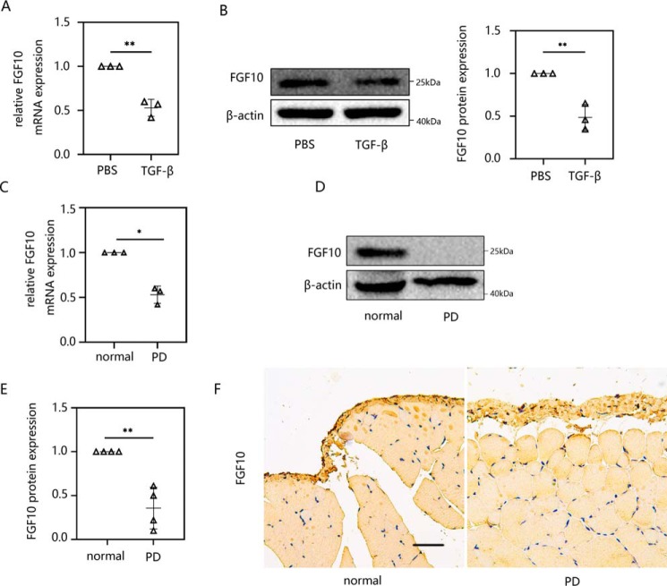
Figure 3.
FGF10 was suppressed in both in vitro and in vivo peritoneal fibrosis models: TGF-β1–induced HMrSV5 cells and PD-treated mice. A and B, HMrSV5 cells were starved for 12 h and then treated with 10 ng/ml TGF-β1. After 24 h, cells were harvested. The expression levels of FGF10 mRNA (A) and protein (B) were evaluated using RT-qPCR and Western blot analysis. C–E, male C57BL/6J mice were intraperitoneally injected daily with 3 ml of 4.25% dextrose PD solution for 4 weeks before parietal and visceral peritoneum tissues were collected. The mRNA (C) and protein level (D and E) of FGF10 was analyzed by RT-qPCR and Western blotting. F, immunohistochemistry was performed to measure the FGF10 response. Data are presented as the mean ± S.D. (error bars) for three independent samples in each group. *, p < 0.05; **, p < 0.01 versus the control group. Scale bar, 50 μm.

