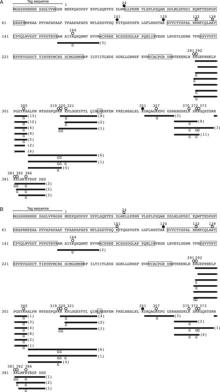
Figure 5.
Ubiquitination sites in hisp53 determined by shotgun MS analysis. A and B, reaction products with WT Ub (A) or with UbK48R (B) were subjected to MS analysis. Peptide fragments with G-G modifications detected by MS analysis are shown as black bars with numbers in parentheses. The numbers indicate the number of fragments detected. The modification sites in the fragments are indicated by G. Peptide fragments not covered by MS analysis are enclosed by open boxes. Lysine residues marked by open circles indicate that ubiquitination was detected, those marked by closed circles indicate that ubiquitination was not detected, and those marked by open squares indicate that the ubiquitination status is unknown.

