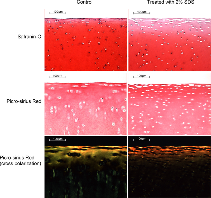
Figure 1.
Photomicrographs of porcine stifle joint cartilage which demonstrate the effect of SDS treatment on tissue cellularity, GAG content (safranin-O), collagen content (picro-sirius red), and collagen organization (picro-sirius red under cross polarization). SDS depleted GAG, particularly in the superficial zone, but had no discernable effect on either collagen content or organization.

