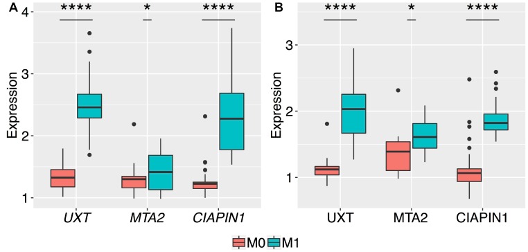
Figure 3. Box plots of the normalized relative expression of UXT, MTA2, and CIAPIN1 genes in the gastric tumor tissues of patients without metastasis (M0) and with metastasis (M1).
The expression levels of the 3 genes were validated by qRT-PCR and western blot in 68 patients, presented with no serosal invasion (T1 and T2). A highly significant increase in the expression of (A) mRNA, and (B) proteins of UXT and CIAPIN1 genes between M0 and M1 stages (**** P < 0.0001). The t-test produced a higher P-value for differences in the mean expression of mRNA and protein of MTA2 as compared to those of other genes (* P = 0.03 and * P = 0.01, respectively). The boxes are drawn from the 75th percentile to the 25th percentile. The horizontal line inside the box represents the median. Vertical lines above and below the box delineate the maximum and minimum values, and the dots show the outliers.

