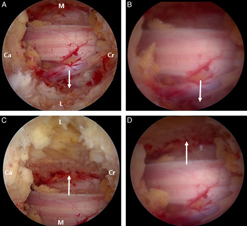
FIGURE 2.

Comparison between the 0-degree and 30-degree arthroscopes. White arrows indicate the lateral recess space. (A) Ipsilateral view with the 30-degree arthroscope. (B) The same view with a 0-degree arthroscope. The contralateral views with a 30-degree (C) and 0-degree arthroscope (D) are shown. Ca indicates caudal; Cr, cranial sides; L, lateral; M, medial.
