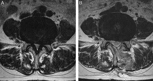
FIGURE 3.

The target spinal level on an axial view of an MRI scan. The patient was a 45-year-old man with grade 3 LCCS who presented with upper gluteal pain radiating to both lower extremities and severe neurogenic claudication with walking ability limited to <100 m. Axial views of L4/L5 in the preoperative state (A) and 1 week after operation (B) are shown. After UBESS, the follow-up MRI scan showed grade 1 LCCS. His clinical symptoms improved and walking capacity increased to 1500 m. LCCS indicates lumbar central canal stenosis; MRI, magnetic resonance imaging; UBESS, unilateral biportal endoscopic spine surgery.
