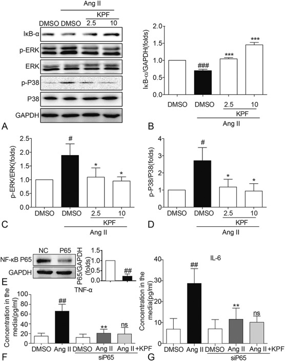
FIGURE 4.

Kaempferol reduced the expression of IκB-α, p-ERK, and p-P38 in Ang II-stimulated cardiac fibroblasts. Cardiac fibroblasts pretreated with 2.5-mM and 10-mM KPF for 24 hours were incubated with 10−8 M Ang II for 24 hours. Western blot analysis was conducted on extracts from cardiac fibroblasts with GAPDH as a loading control (A). The normalized optical density of IκB-α (B), p-ERK (C), and p-P38 (D) in mean ± SE was from 3 independent experiments, respectively. E, Western blot analysis of P65 after siRNA transfection in cardiac fibroblasts. F, G, Effect of P65 silencing and KPF on Ang II-induced TNF-α (F) and IL-6 (G) mRNA levels (# vs. DMSO group; * vs. DMSO + Ang II group; ns, no significant, vs. siP65 + Ang II group; # and *P < 0.05, ## and **P < 0.01, ### and ***P < 0.001). DMSO, dimethylsulfoxide; GAPDH, glyceraldehyde‐3‐phosphate dehydrogenase.
