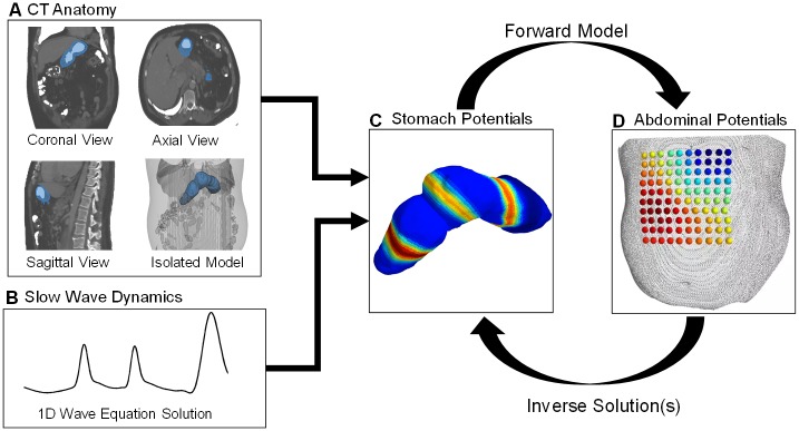
Fig 1. Process workflow.
A) Using the 3 standard CT views, a 3D model of the stomach is extracted. B) We develop a spatially inhomogeneous solution to the 1D wave equation, pertaining to propagation down the organoaxial direction of the stomach, with region-specific amplitudes and speeds based upon recent findings in the literature from invasive human recordings. C) The 1D wave equation solution is mapped to the 3D model to generate the dynamic dipole moment solution. D) We solve a forward model to generate the dynamic simulated observations and inject additive measurement noise.

