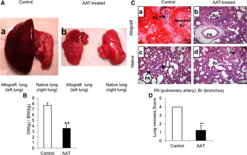
FIGURE 3.

Treatment with AAT attenuated lung allograft injury and necrosis. To investigate the potential therapeutic benefit of AAT on posttransplantation IRI, the orthotopic left single lung transplant was performed between Lewis (donor) and SD (recipient) rats. A, Representative gross images of lungs in the control rats (panel a) and treatment group (panel b). B, Treatment with AAT significantly reduced the GW:BW ratio in the treatment group compared with the control group. Data represent the mean plus SEM; **P < 0.01, (n = 6 rats in control group and 5 rats in the AAT treatment group). C, Histologic examination (H&E stained, ×200 magnification) showed an extensive necrosis of lung allografts in the control group (panel a) in comparison to lung allografts in the treatment group (panel b) and native lungs (panels c and d) on day 8 posttransplantation. D, Semiquantitative lung necrosis scoring was performed using a 5-point scale according to the percent involvement of necrosis in each section. The mean percent necrosis score was significantly less in the AAT treatment group in comparison to the control group. Data represent the mean plus SEM; n = 6 in control group and n = 5 in the AAT-treated group. AAT, alpha-1 antitrypsin; GW/BW, allograft weight/body weight; H&E, hematoxylin and eosin; IRI, ischemia-reperfusion injury; SD, Sprague-Dawley; SEM, standard error of the mean.
