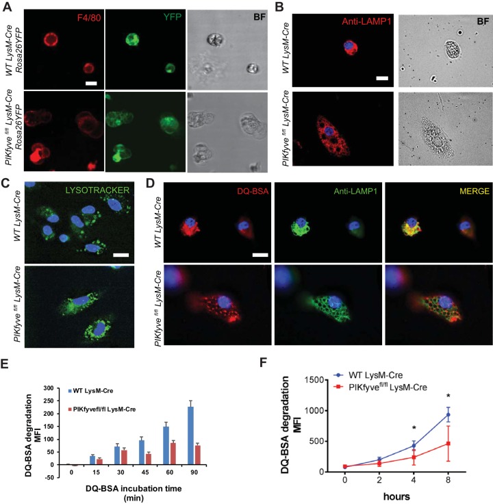
FIG 4.
PIKfyve modulates lysosomal morphology and degradative function in macrophages. (A) Live-cell fluorescence microscopy images of spleen macrophages isolated and stained with F4/80 antibody. The intrinsic YFP expression is driven by LysM-Cre. Note the presence of multiple cytoplasmic vacuoles of various sizes in the macrophages of a PIKfyvefl/fl LysM-Cre Rosa26YFP mouse. BF, bright-field. (B) Immunofluorescence images of bone marrow-derived macrophages stained with anti-LAMP1 antibody. (C) Images of bone marrow-derived macrophages incubated with LysoTracker for 20 min. Note the accumulation of LysoTracker in the acidic endolysosomes. (D) Immunofluorescence images of bone marrow-derived macrophages incubated with DQ BSA for 60 min and costained with anti-LAMP1 antibody. (E) Quantification of DQ BSA degradation over 90 min visualized by live microscopy. MFI, mean fluorescence intensity. (F) Proteolytic DQ BSA degradation over 8 h in the F4/80+ spleen macrophages as measured by increasing fluorescence of quenched dye on a spectrophotometer. Statistical analysis was performed using unpaired two-tailed Student’s t test (*, P < 0.05). All error bars indicate SEM. Scale bars: 10 μm.

