
Figure 6.
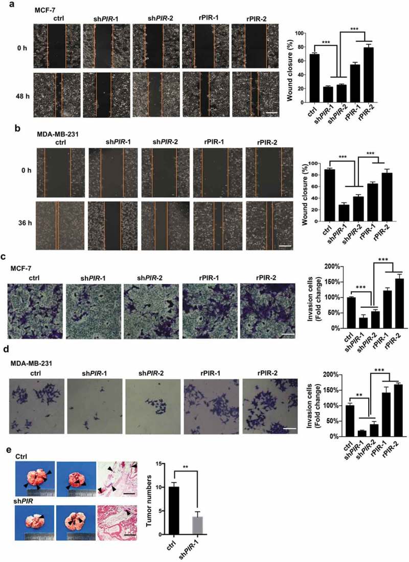
PIR promotes tumor cell migration, invasion and metastasis. (a,b) The same MCF7 (a) and MDA-MB-231 (b) cell lines with PIR knocked down and re-expressed as those used in Figure 2 were plated. After 24 h of plating, artificial wound was created by a 200 ml pipette tip, followed by determination of wound closure under microscope after another 48 h for MCF7 and 36 h for MDA-MB-231 cells. Left, representative morphologies; right, statistic results shown as means±SEM of three independent experiments (***P< 0.001, unpaired Student’s t-test). The scale bars represent 100 mm. (c,d) The transwell assay were performed with the same MCF7 (c) and MDA-MB-231 (d) cells as those used in wound healing assay. Left, representative morphologies; right, statistic results shown as means±SEM of three independent experiments (*P< 0.05, **P< 0.01,***P< 0.001, unpaired Student’s t-test). The scale bars represent 50 mm. (e) MCF7 cells (1´106 in 100 μl PBS) with PIR stably knocked down were injected into the four-week-old male BALB/c nude mice via tail veins. The mice were sacrificed after 6 weeks and pictures of metastatic lungs were taken. The scale bars represent 1 mm.