. 2019 Oct 3;8:e48408. doi: 10.7554/eLife.48408

Figure 7. Functional segregation of in vivo electrophysiological properties mSN DA neurons with distinct axonal projections.

(A1, B1) Spontaneous in vivo extracellular single-unit activities of a representative DS-projecting DA neuron located in the mSN (A1) and a representative lNAcc-projecting DA neuron located in the mSN (B1), shown as 10 s of original recording traces (scale bar: 0.2 mV, 1 s). Note the differences in bursting. (A2, B2) 30 s raster plots (scale bar: 1 s). (A3, B3) ISI-distributions. Note the presence of ISIs below 80 ms and 160 ms indicating bursts in B3 in contrast to A3. Inset, averaged AP waveform showing biphasic extracellular action potentials in high resolution (scale bar: 0.2 mV, 1 ms). (A4, B4) Confocal images of retrogradely traced, extracellularly recorded and juxtacellularly labelled DA neurons, the location of the neurons is displayed in the bottom right images. (C) Anatomical mapping of all retrogradely traced, extracellularly recorded and juxtacellularly labelled neurons (projected to bregma −3.16 mm; DS-lSN in blue, lNAcc-mSN in magenta). Inset, FG-injection sites in DMS, DLS and lNAcc (FG in blue, TH in green). (D) Cumulative SFB distribution histograms (dotted line at SFB = 5% threshold) and bar graphs of SFB contingencies (% of neurons > and < 5% SFB) showing significant differences in burstiness. (E) Cumulative distribution histograms and bar graphs of pauses per minute showing significant differences. (F) Scatter dot-plots showing significant differences in kurtosis and skewness of the ISI-distributions. (G) Linear discriminant analysis of DA mSN neurons projecting either to DLS/DMS or lNAcc. 70.8% of neurons were classified correctly based on skewness of the ISI-distribution, AP duration, repolarization speed and the precision of spiking (log(σ2)) when randomly bisecting the data 1000 times. When the whole data set was used, 84.4% of neurons were classified correctly. (Right Picture) Mapping of correctly- vs incorrectly-classified DA mSN neurons projecting to DS or lNAcc.

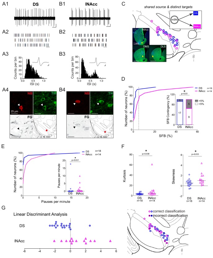
Figure 7.

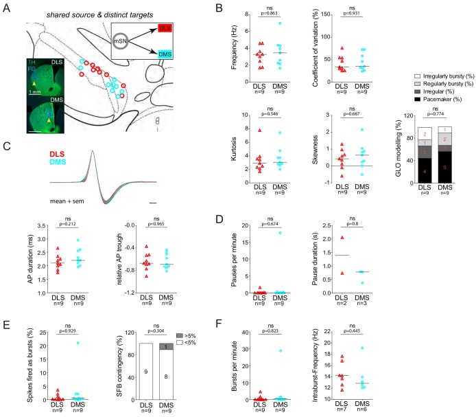
Figure 7—figure supplement 1. DLS and DMS-projecting DA neurons located in the mSN do not exhibit significantly different in vivo firing properties.

Figure 7—figure supplement 1.

(A) Anatomical mapping of all extracellularly recorded and juxtacellularly labelled neurons (projected to bregma −3.16 mm; DLS in red, DMS in cyan). Inset, FG-injection sites in the DLS and DMS (FG in blue, TH in green). (B–F) Scatter dot-plots and bar graphs (line at median) showing no significant differences in firing frequency (Hz), coefficient of variation (%), kurtosis and skewness of the ISI-distributions, GLO-based firing pattern (all B), normalized AP waveform, AP duration (ms), relative AP trough (all C), pauses per minute, pause duration (s) (both D), SFB (%), SFB contingency (% of neurons > and < 5% SFB) (both E), bursts per minute, intraburst-frequency (Hz) (both F).
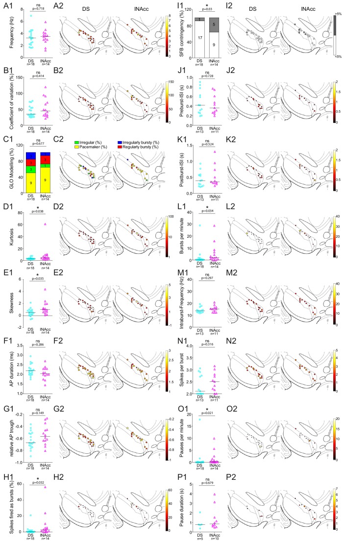
Figure 7—figure supplement 2. Comparison of in vivo firing properties of DS and lNAcc-projecting DA mSN neurons.

Figure 7—figure supplement 2.

(A1–P1) Scatter dot-plots (line at median) and bar graphs showing significant differences in kurtosis, skewness, SFB (%), SFB contingency (% of >and < 5%), bursts per minute, pauses per minute. (A2–P2) Feature maps of respective parameters.
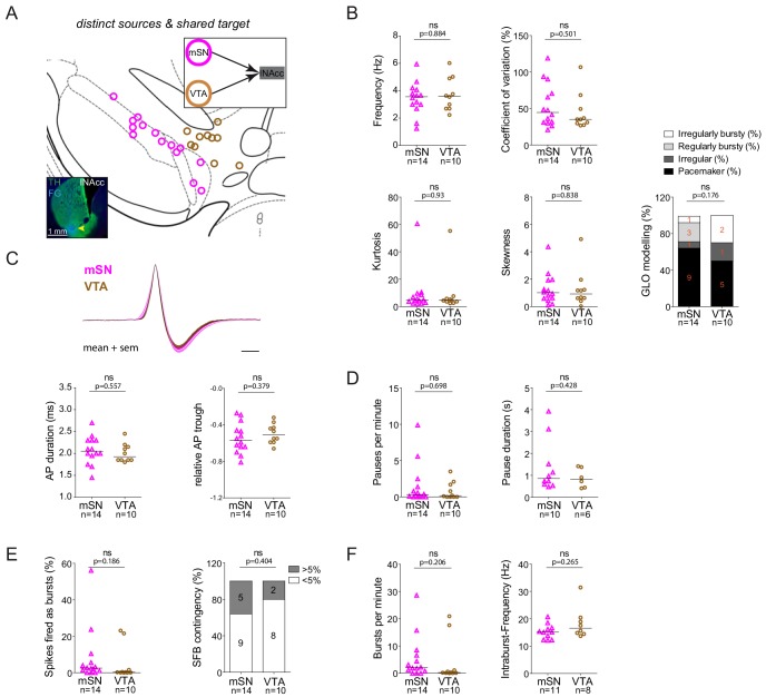
Figure 7—figure supplement 3. lNAcc-projecting DA neurons located either in mSN or VTA do not exhibit significantly different in vivo firing properties.

Figure 7—figure supplement 3.

(A) Anatomical mapping of all extracellularly recorded and juxtacellularly labelled neurons (projected to bregma −3.16 mm; mSN in magenta, VTA in brown). Inset, FG-injection site in the DLS (FG in blue, TH in green). (B–F) Scatter dot-plots (line at median) and bar graphs showing no significant differences in firing frequency(Hz), coefficient of variation (%), kurtosis and skewness of the ISI-distributions, GLO-based firing pattern (all B), normalized AP waveform, AP duration (ms), relative AP trough (all C), pauses per minute, pause duration (s) (both D), SFB (%), SFB contingency (% of neurons > and < 5% SFB) (both E), bursts per minute, intraburst-frequency (Hz) (both F).