Figure 1.

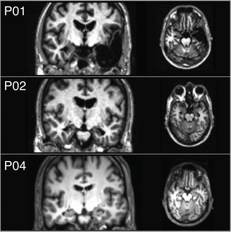
Coronal and axial T1-weighted magnetic resonance images depict lesions for patients P01, P02, and P04 (no scans were available for P03). The left side of the brain is displayed on the right side of the image.

Coronal and axial T1-weighted magnetic resonance images depict lesions for patients P01, P02, and P04 (no scans were available for P03). The left side of the brain is displayed on the right side of the image.