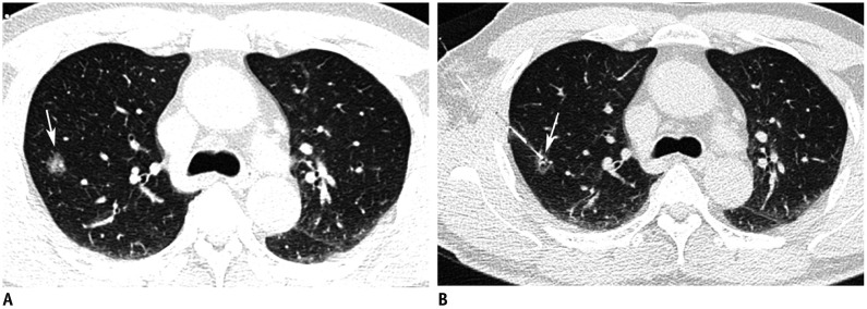
Fig. 1. Preoperative hook-wire localization for part-solid nodule in right upper lobe in 59-year-old man.
A. Axial CT image with lung window shows 15-mm part-solid nodule (arrow) in right upper lobe. B. Post-procedure axial CT image with lung window shows that horn of hook-wire is positioned in target nodule (arrow).

