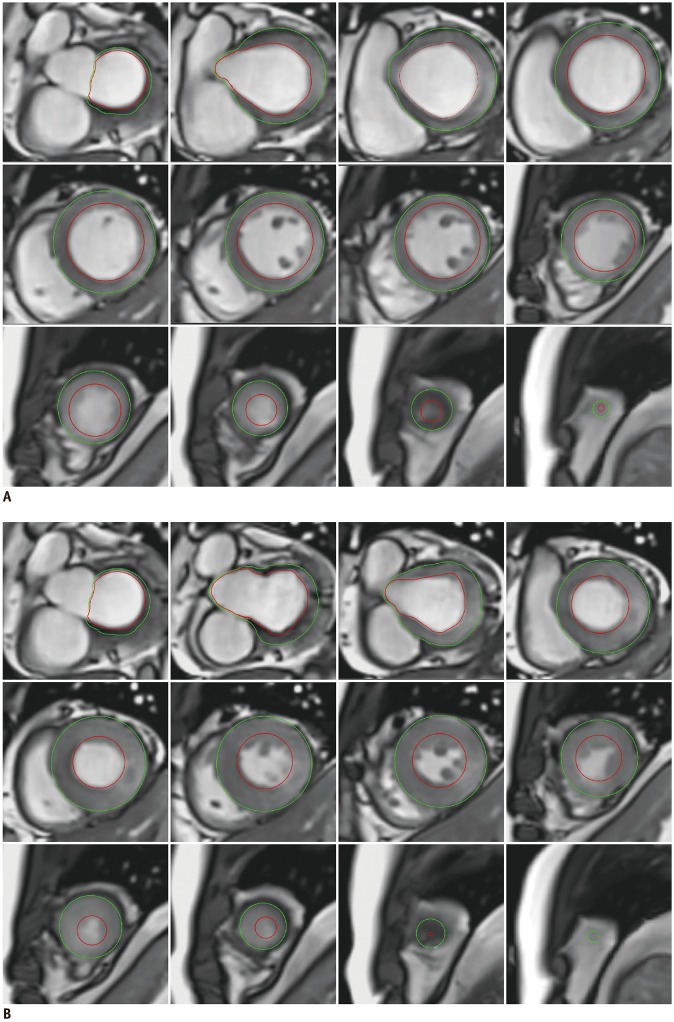
Fig. 2. LV quantitative assessment.
For LV quantitative assessment, stack of short-axis slices containing entire LV is required, and endocardial (red) and epicardial (green) contours should be drawn in both diastole (A) and systole (B) phases. Inclusion or exclusion of papillary muscles should be mentioned. Note that papillary muscles are excluded in this example.

