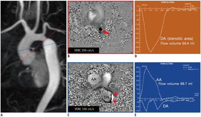
Fig. 6. Case of 31-year-old man with coarctation of aorta.
A. Contrast-enhanced MR angiography showing coarctation of aorta at isthmic portion of aorta. B. Velocity map crossing stenotic lesion showing flow acceleration (arrow) at coarctation site (Supplementary Movie 1). C. Velocity map at level of AA. Because of low VENC value of 150 cm/s, aliasing artifact (arrowheads) occurred along wall of DA, just at distal portion of coarctation site. D, E. Time-flow volume curve maps showing change in flow volume along cardiac cycle. AA = ascending aorta, DA = descending aorta, VENC = velocity encoding

