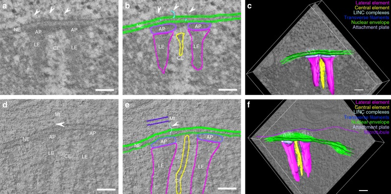
Fig. 4.
Segmentation for 3D model generation of telomere attachment sites. a, d Single virtual section of a reconstructed tomogram of a telomere attachment site without a microtubule (a) and with a microtubule running parallel to the frontal view of the synaptonemal complex (d). b, e Respective manual segmentation of the virtual sections of a and d. c, f Resulting 3D models of telomere attachment sites from the combination of all individual segmentations. LE: lateral element, CE: central element, AP: attachment plate, Ch: Chromatin, NE: nuclear envelope, Mt: microtubule. Arrowheads indicate LINC complexes associated with the attachment sites. Scale bars: 100 nm

