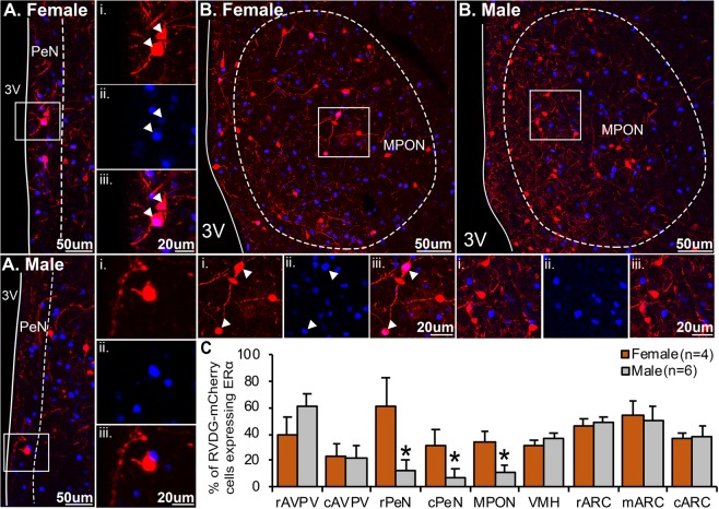
Figure 6.
Sexually differentiated input to KNDy neurons from estrogen-sensitive cells in the periventricular nucleus and the medial preoptic area. Confocal images of mCherry-immunoreactive (ir) (red) afferent populations to KNDy neurons and estrogen receptor alpha (ERα)-positive nuclei (blue) in the periventricular nucleus (PeN, A) and medial preoptic nucleus (MPON, B) of male and female mice. High magnification images of mCherry-positive neurons (i), ERα positive nuclei (ii) and merged images from corresponding boxes in (A,B). (C) Histogram depicting the mean ± SEM percentage of afferent populations to KNDy mCherry-ir cells colocalized with ERα in the rostral (rAVPV) and caudal (AVPV) anteroventral periventricular nucleus, rostral (rPeN) and caudal (cPeN) PeN, ventromedial hypothalamus (VMH), and rostral (rARC), middle (mARC) or caudal (cARC) arcuate nucleus. The percentage of afferents to KNDy mCherry-ir cells colocalized with ERα is significantly higher in female mice compared to male mice in the rPeN, cPeN and MPON. No significant difference is detected in the rAVPV, cAVPV, VMH, rARC, mARC or cARC. 3 V, third ventricle. Student’s t-test.

