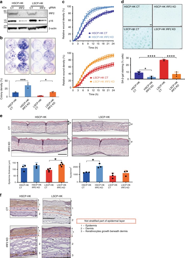
Fig. 7.
IRF2 loss of function in LPSC-HK restores stem cell potential. a Effect of CRISPR–Cas9 KD of IRF2 in HSCP-HK and LSCP-HK cells on IRF2 and p16 INK4A protein expression as measured by Western blot using β-actin as a loading control. b Clonogenic assay comparing IRF2 KD in HSCP-HKs (left panel) versus LSCP-HK (right panel) and quantification of colony forming potential as a function of cell density. Colony density was measured in % area (means ± S.D. of n = 3 biological replicates). *p < 0.05, ***p < 0.001 (One-way ANOVA with Holm–Šídák multiple comparisons test). c Keratinocyte migration assay comparing IRF2 KD in HSCP-HK (top panel) versus LSCP-HK (bottom panel). d Beta-galactosidase (SA-β-gal) staining as a marker of senescence upon IRF2 KD in HSCP-HK versus LSCP-HK cells. SA-β-gal staining was quantified as % of cells with positive staining (means ± S.D. of n = 3 biological replicates). *p < 0.05, ****p < 0.0001. (One-way ANOVA with Holm–Šídák multiple comparisons test). Scale bar = 200 μm. e Quantification of epidermis formation upon IRF2 KD in HSCP-HK cells versus LSCP-HK cells. * indicates p < 0.05 (One-way ANOVA with Holm–Šídák multiple comparisons test), # indicates the epidermal layer. Epidermis thickness (μm) was measured for each condition at 3 sites within the construct (means ± S.D. of n = 4 biological replicates). The number of DAPI-stained nuclei were counted per mm2 for each condition. Scale bar = 167 μm. f Immunocytochemical staining for Keratin 10 in human dermo-epidermal 3D model comparing IRF2 KD in HSCP-HK versus LSCP-HK. Keratin 10 is used as an epidermal differentiation marker expressed only by keratinocytes in suprabasal epidermal layers in human skin. Keratin 10 staining reflects differentiation patterns within 3D skin constructs where the presence and structure of a basal (stem cell) layer does not stain. For clarity, epidermis is labeled as layer 1, dermis as layer 2 and keratinocytes growing beneath the dermis are labeled as layer 3. Red dotted lines illustrate the extent of the unstained basal layer within the epidermis. While all human skin constructs maintain an unstained (undifferentiated) basal layer, differences between conditions are apparent. Scale bar = 128 μm

