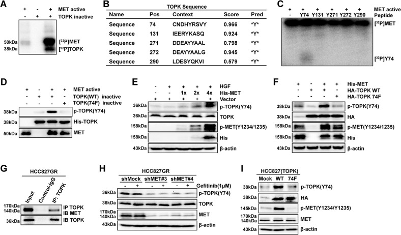
Fig. 4. MET phosphorylates TOPK at the Y74 site in vitro and ex vivo.
a Active MET phosphorylated inactive TOPK-WT in vitro in the presence of [γ-32P] ATP as visualized by autoradiography. b Potential phosphorylated Tyr sites of TOPK were predicted by the NetPhos 3.0 software program. c Synthesized peptides containing potential Tyr b sites were used as substrates in an in vitro kinase assay with active MET in the presence of [γ-32P] ATP and the results were visualized by autoradiography. d Active MET phosphorylated inactive TOPK-WT or 74F in vitro in the presence of ATP. Then, the samples were analyzed by western blotting. e MET promoted the phosphorylation of TOPK in HEK293T cells induced by HGF after transfection with His-MET in a dose-dependent manner (HGF 40 ng/ml; 15 min). f His-MET was cotransfected in HEK293T cells with wild-type TOPK (HA-TOPK WT) or Y74-mutated TOPK (HA-TOPK 74F). g TOPK bound with endogenous MET of HCC827GR cells by IP assay. h The stable shMock and shMET in HCC827GR cells were treated with gefitinib (1 μΜ) for 6 h. i HCC827 cells stably overexpressing Mock, HA-TOPK WT, and HA-TOPK 74F were cultured and analyzed by western blotting. The above samples were collected and analyzed by western blotting. Data are representative of results from triplicate experiments

