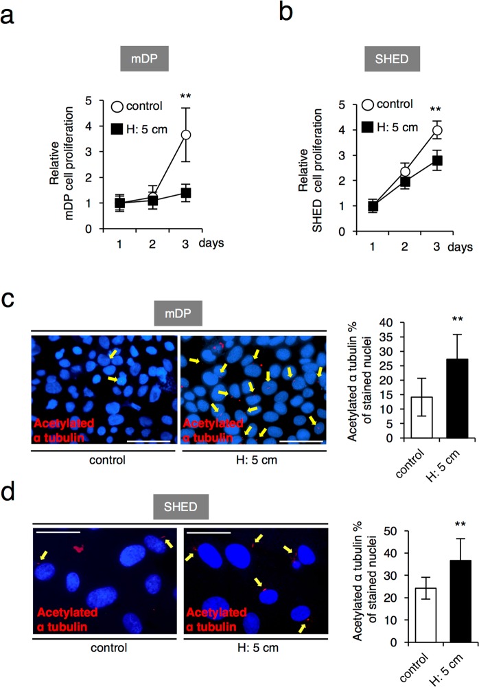
Figure 2.
Sustained HP inhibits cell proliferation and induces ciliogenesis in SHED. (a) Cell proliferation analysis by a cell counting method in mDP cells (a) and SHED (b). The cells were cultured with or without the HP by the medium height of 5 cm (H: 5cm) for 24, 48 and 72 hrs. (c,d) Immunostaining of acetylated α-tubulin in mDP cells (c), Scale bar: 50 μm; and SHED (d), Scale bar: 25 μm. The cells were cultured with or without the HP by the medium height of 5 cm (H: 5 cm) for 6 hrs, and then immunostaining was perfomed for acetylated α-tubulin. The acetylated α-tubulin-positive cells were counted in twenty randomly selected fields of view under an inverted microscope (20X magnification). The bar graph shows the percentage of α-tubulin-positive cells of total nuclear-stained cells. Red, acetylated α-tubulin; blue, DAPI. The data presented in (a,b) is a representative of three independent experiments showed similar results. (c,d) Represent the mean (±standard deviation, SD) of three independent experiments, and each performed in triplicate. Statistical analysis was performed using analysis of variance (**p < 0.01).

