. 2019 Oct 4;33(5):436–445. doi: 10.3341/kjo.2019.0036

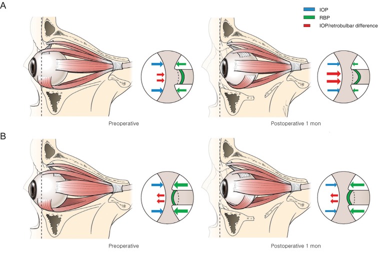
Fig. 3. Schematic illustration of the change in lamina cribrosa depth after orbital decompression, comparing the fat-dominant group and the muscle-dominant group. (A) In the fat-dominant group, at baseline, retrobulbar pressure (RBP) was lower than that of the muscle-dominant group. Therefore, the translaminar pressure difference moves to the posterior surface of the lamina cribrosa. After orbital decompression, the RBP decreased, and the net translaminar pressure difference increased toward the same direction. (B) In the muscle-dominant group, at baseline, the RBP was relatively larger than that of the fat-dominant group, and the translaminar pressure difference was directed toward the anterior surface of the lamina cribrosa. After surgery, intraocular pressure (IOP) and RBP decreased slightly at the same time. Therefore, there was no definite change in translaminar pressure difference. Cerebrospinal fluid pressure is not indicated in this diagram under the assumption that it did not change after the surgery.

Fig. 3