Fig. 2.
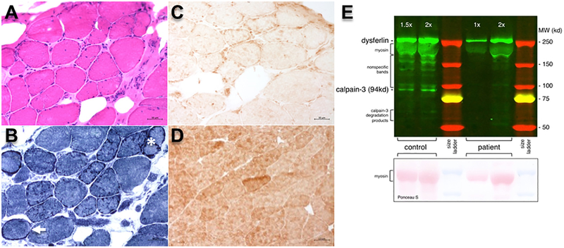
Muscle histological and western blot findings. Biceps femoris muscle biopsy of the proband showing muscle fiber variation and an increase in fibrous and fatty connective tissue (A, hematoxylin and eosin). NADH dehydrogenase reacted sections (B) show numerous lobulated fibers (asterisk) or ring fibers (arrowhead). (C) Calpain-3 immunoreactivity (anti-calpain-3 2C4 antibodies) is reduced in the proband. (D) A normal muscle section with preserved calpain-3 immunoreactivity is shown for comparison. (E) Control muscle was compared to the muscle biopsy from the proband. While dysferlin appears normal in size and amount for the proband, full-length (94kd) calpain-3 and calpain-3 degradation products are greatly reduced. The antibodies used for Western blotting were Hamlet (anti-dysferlin) and 12A2 (anti–calpain-3), both purchased from Leica Biosystems.

