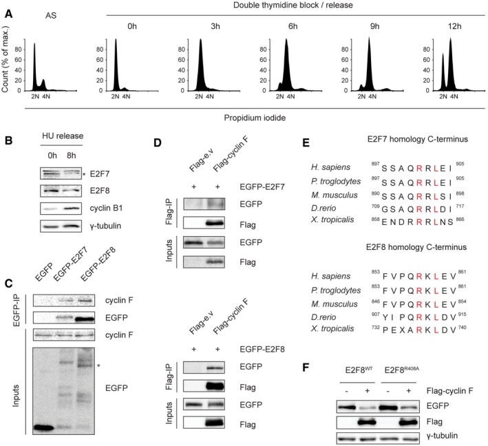
Figure EV1. Cyclin F binds to E2F7 and E2F8.

- Cell cycle analysis of the double thymidine block and release experiment shown in Fig 1A. HeLa cells were synchronized by double thymidine block and then released in fresh medium. Asynchronized (AS) and synchronized cells were harvested at the indicated time points for propidium iodide staining and flow cytometry analysis.
- E2F7 and E2F8 are degraded during G2/M phases. HeLa cells were treated with hydroxyurea (HU, 2 mM) for 16 h to arrest cells at G1/S border. Then, HU was removed and cells were released into fresh medium. Protein samples were harvested at the onset of release and 8 h after release. E2F7/8 levels were measured by immunoblotting. Protein expression of cyclin B1 was used as a marker for G2 or M cell cycle progression, and γ‐tubulin was used as loading control. Asterisk indicates the specific band of E2F7 detection.
- Immunoprecipitation shows that cyclin F physically interacts with E2F7/8 in vitro. HEK293 cells were transiently transfected with either EGFP‐tagged empty vector (EGFP), EGFP‐tagged E2F7 or EGFP‐tagged E2F8. MG132 was added to the cells 5 h before harvesting at 48 h post‐transfection. Cells were harvested and lysed for immunoprecipitation using GFP resin. Asterisk indicates the E2F8‐specific band.
- Reciprocal IP demonstrated the bindings between cyclin F and E2F7/8. HEK cells were transfected with Flag‐tagged empty vector or cyclin F, together with GFP‐tagged E2F7 or E2F8. MG132 was added to cells 5 h before harvesting, and co‐IP was performed using Flag resin.
- Homology of atypical E2Fs at their C‐terminus shows that the cyclin F‐binding motifs are conserved among different species.
- Wild‐type or R408A mutant of EGFP‐tagged E2F8 was co‐transfected with either empty vector or Flag‐tagged cyclin F in HEK293 cells. Nocodazole was added to cells 8 h before harvest. Forty‐eight hours after transfection, cells were collected and lysed for immunoblotting.
Source data are available online for this figure.
