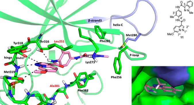
Figure 3.
X-ray structure of LCK and 11. Hydrogen bonds and favorable interactions are denoted with dashes. The carbons and ribbon representation of LCK are colored green except for the loop between β-strand3 and helix-C which is colored blue. The carbons of 11 are colored pink, oxygen is red, and nitrogen is blue. Residues in the binding pocket that differ between LCK and CSK have red labels. Bottom right: Close-up of the surface of LCK around the indazole. The surface of LCK is colored green except for the loop between the β-strand3 and helix-C which is colored blue.

